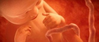
The first smile of a one-month-old baby is the unforgettable joy of its parents. Although she is rare and uncertain, she is very significant. At 2 months, the smile becomes more and more conscious; it appears when an adult addresses the baby. At 3 months, a smile already signifies joy and is accompanied by animated movements of the arms and legs, which certainly delights adults. When a baby smiles or laughs in his sleep, it causes anxiety.
Smile of a newborn
Why do newborns smile in their sleep?
For newborn children, everything in this world is so new and unfamiliar, all the time he learns something unusual, gains impressions and positive emotions. Why do newborns smile in their sleep? But that is precisely why they smile. All emotions and impressions from the day he experienced are reflected in his dreams. But it's important to remember that both positive and negative events affect sleep in the same way . Therefore, doctors advise not to overload the baby with a variety of entertainment at first. But if your newborn baby smiles in his sleep, then the past day has definitely brought him a lot of positive emotions.
Another reason why a newborn smiles in his sleep may be a change in sleep phases . There are fast and slow sleep phases. When changing from one phase to another, the baby may smile, laugh, move his arms and even mutter. This is believed to be normal.
Another slightly strange answer to the question “Why do newborns smile in their sleep?” is that angels seem to fly to the baby and play with him. It is believed that in this case the child should not be woken up. The above versions are not a cause for concern for parents.
Actions of parents of a laughing child
Laughter in a dream should be treated as a positive phenomenon. Laughter is a positive and even useful emotion. Children who giggle do not feel afraid at night, unlike those who scream and cry. The fun for these kids doesn’t end even during sleep.
Hiccups in newborns after feeding - what to do?
If a child doesn’t just giggle for a couple of minutes from time to time, but laughs out loud long enough and often, it’s worth thinking about the state of his nervous system. Perhaps he has a mobile psyche, he reacts very violently to events that impress him. Such kids need to be protected as much as possible from unnecessary emotional stress and be extremely careful with ideas such as:
- invite a clown to your birthday party;
- watch fireworks in close proximity to it;
- turn on a noisy household appliance without discussing it with the child in advance (or without sending him for a walk while using loud equipment);
- watch films in the presence of a child that are not age-appropriate and contain scenes of violence or vivid images of negative characters.
Important! Overly emotional children need to be protected. Be sure to discuss their experiences with them. Hidden fears or insecurities are not always expressed through nightmares. Sometimes the psyche responds to nervous excitement by laughing at night.
If your baby often laughs at night, it is important to end the day in a certain way and properly prepare for bed:
- three hours before bedtime, exclude TV, tablet and computer;
- The background sound of calm children's kind songs is allowed;
- the child can engage in quiet activities: draw, sculpt, play with sand;
- immediately before bed, take a bath; if we are talking about a child who already knows how to sit and play, then give him two vessels for pouring water;
- after the bath, slowly massage your arms, legs and back using baby cream or pharmaceutical oil;
- Tea with chamomile or mint, which has a calming effect, will not be superfluous;
- the light after the bath should be diffused, dim, not bright;
- When the child lies down in a clean bed, they sing him a lullaby or read a good fairy tale.
Bedtime story
It is very important for receptive children to provide a smooth and long transition from an active day to a quiet evening. With children who can talk, it is important to discuss their day before going to bed. Trusting, open communication is useful both for forming strong family relationships and for helping the psyche sort out difficult moments.
First, the child is asked about what he did not like about his day. If he finds it difficult to answer, you can suggest, for example, the apple turned out to be sour or he didn’t want to leave the walk. In order for the child not to feel alone with the problem (it was not possible to inflate the balloon), an adult can tell a similar situation from his childhood to share his experience or explain that at this age it is not easy to cope with the task. Then the child is asked what he liked most today and why. On this positive note, the child should fall asleep.
Some resources claim that laughter in a dream is a sign of terrible pathologies of the central nervous system or a symptom of a tumor in the brain. We can definitely say that laughter without any special accompanying symptoms is not scary. In case of serious illnesses, a person has other problems, both with mental and physical health, which will indicate the presence of the disease before nightly laughter.
For a restful child's sleep, it is enough to give the baby the opportunity to spend as much energy as possible during the day, and then go to bed with a certain ritual. When a child laughs, do not worry - this is normal. Laughter is most often a manifestation of joy and delight. Thanks to him, we can consider that the day was spent as positively and educationally as possible.
What does a baby's smile in a dream mean?
If you are still concerned that your child often smiles or laughs in his sleep, you can contact a specialist to dispel all unnecessary fears. Sometimes the baby may be prescribed to drink soothing herbal infusions before bed.
Remember that nighttime sleep is very important for a child. After all, this is precisely the time when the body grows, rests, and carries out vital processes. Therefore, it is important that nothing disturbs or interferes with the baby, so that the sleeping conditions are as comfortable as possible. To do this, try to adhere to the following tips:
- Ventilate the room where your child sleeps before he goes to sleep.
- Before going to bed, you need to exclude active games, watching cartoons on the computer or TV (it’s better to read a book or just talk quietly).
- Before going to bed, it is advisable to take an evening walk; this has a positive effect on sleep.
Before bed rituals
Of course, these are not all the reasons for poor sleep in newborns. Often, restlessness, anxiety and crying can be a reaction to new strong emotions or uncomfortable clothing. But there is a universal recipe for combating insomnia in newborns - these are bedtime rituals:
- Always put your child to bed at the same time. A daily routine helps relieve anxiety in a baby.
- Choose clothes made from natural fabrics with soft and wide elastic bands.
- Give your baby a light relaxing massage.
- Sing a lullaby. Mom’s calm, even voice is the best sleeping pill.
- Lie down with your child. Pet him. Body contact in the first six months of life is not a whim, but a necessity.
Share on social media networks
How to ensure healthy sleep for a newborn
In the first time after birth, it is very difficult for parents to create favorable conditions for the baby. The child cannot fall asleep and wake up on command.
A baby who has recently been born is just beginning to adapt to life in this world, just getting used to and adapting to the environment. Therefore, you need to try to create the most comfortable and cozy conditions for sleep and development.
Babies are called newborns up to a month old. At first they sleep a lot, up to 20 hours a day. The baby sleeps in the so-called “frog” position with slightly bent legs and arms spread to the sides.
If the baby sleeps for a long time, do not sound the alarm ahead of time. The older the child is, the less time his sleep will last. For example, already at three months the baby will sleep for about 15 hours . Sleep in young children does not last long, about 3-4 hours . Then comes the period of feeding, playing, changing diapers, etc. Experts advise accustoming your baby to a certain schedule, then it will be easier for both mother and baby in the future.
If nothing works out with the schedule, don’t rush to get upset, everything will work out . When your baby wants to sleep, you will understand this by his behavior: he will begin to rub his eyes, dark circles appear under them. Remember that you should not train your baby to fall asleep when you breastfeed or rock him to sleep.
Try to teach your child to be independent from an early age. Put the child to bed half asleep, sing lullabies that soothe thanks to the quiet and gentle voice of the mother, turn off the bright lights. Remove extra pillows, blankets, etc. from the crib, as this will disturb and negatively affect your baby's sleep. In addition, it is recommended that babies under one year of age sleep without a pillow, so as not to harm the spine.
It is best if the baby sleeps on a firm mattress. For sleep, choose comfortable, soft clothes so that they do not cause discomfort.
When to be wary
Sometimes, when the baby is sleeping, some mysterious phenomena occur. They alarm parents and make them wary. And not in vain.
Here are two main signs that require attention:
We recommend reading! Click on the link:
Common reasons why a child does not sleep at night and cries
- crying that suddenly appears and disappears just as abruptly;
- unexpected and abruptly ending laughter.
The common features of these two phenomena are an abrupt onset and an abrupt end, and the fact that the baby at these moments does not react to others. It is impossible to calm him down.
Isolated cases do not pose a threat. Perhaps the baby simply “went out for a bit”, and the nervous system thus received a release. It is worse if attacks of sudden crying or laughter are repeated frequently and regularly. In this case, show the child to a neurologist. In combination with restless behavior at other times, they may indicate developmental delay, hyperactivity syndrome and some other problems with the central nervous system.
Even a healthy child can laugh and laugh in his sleep. Such laughter is usually preceded by a smile, then light chuckles. Only then does the baby begin to laugh at full strength. This is a completely natural phenomenon. The baby develops normally, eats well, and has deep sleep phases.
Crying and laughter that suddenly appear and disappear just as abruptly should alert the mother. This is an alarm bell indicating some problems with the central nervous system. A pediatrician and a neurologist will help you overcome difficulties.
Why do newborns startle in their sleep?
Very often, in their sleep or when falling asleep, babies flinch, which is very frightening and alarming for mothers. Don't panic, there's nothing wrong with this, this phenomenon will disappear over time. The baby is just getting used to the new world, getting used to new conditions. By the way, premature babies flinch much more often.
So why do newborns flinch?
- The child is tired. You should not play active games with your baby before bedtime. Tired and overtired after this, the child sleeps poorly and shudders in his sleep.
- Colic . Very often, three-week-old babies have tummy problems because their digestive system is just adjusting to normal functioning. That is why at first nursing mothers actually go on a diet, because many foods eaten by the mother can have a bad effect on the child’s condition. If your baby has severe sleep problems that involve startling, consult your doctor for advice.
- Fever . Babies have a very weak immune system, so at first there is a high risk of contracting an infectious disease. If the temperature is really elevated, you need to contact your pediatrician.
- Loud sounds. At night, the baby should be surrounded by peace and quiet. Try to create a cozy atmosphere, pat your baby on the head so that he feels your presence and is calm.
When to ask for help
Now let's look at cases in which your baby needs the help of a specialist. The most widespread insomnia is difficulty falling asleep, as well as regular awakenings. There are the following causes of insomnia in children:
- Secondary – occurs as a consequence of any disease. For example, insomnia in children can occur due to fever or abdominal pain.
- Primary – not associated with any diseases. The main cause of insomnia in children in this case is the behavior of parents or the child during sleep.
Secondary causes of insomnia in children are eliminated by treating the diseases that cause it. Primary – by adjusting behavior patterns during the period of falling asleep. Let's look at how to do this.
Newborn groans in his sleep
Very often the baby grunts if he is constipated . If your child really experiences constipation frequently, it is best to consult a doctor. Some mothers try to administer enemas on their own or give their children laxatives, but the main thing in this matter is not to abuse it. In addition, colic can also cause grunting.
Gases produced during meals press on the intestinal walls, thereby causing pain. To prevent your baby from having painful sensations in the tummy area, give him a special massage and carry him after eating in an upright position so that the air comes out of the esophagus . If the problem is really serious and nothing helps, seek help from your pediatrician.
Features of a child's sleep
Birth and exposure to unknown conditions is a very tiring task for any baby. This is what explains such a long duration of sleep in babies in the first months of life, compared to adults.
And in general, infants sleep completely differently than their parents. The baby's deep sleep phase begins in about half an hour. It does not last long, and after a while it is replaced by the stage of shallow sleep. Its duration is much longer, from which we can conclude that newborns sleep lightly most of the time. For this reason, their sleep can be considered very sensitive, and babies themselves can be considered extremely susceptible to external stimuli.
Often during the superficial phase, young children show various emotions: they may cry, grunt, or, on the contrary, smile and even laugh.
By paying close attention to a child’s behavior during sleep, one can judge his physical and psychological state. For example, by crying, a baby is able to show that he is bothered by intestinal colic, and a groaning child is often constipated. In turn, a smile, according to most researchers, indicates a favorable emotional background and the absence of any disturbances in the functioning of the child’s body.
Thus, for the most part, the shallow sleep of newborns, replete with various kinds of manifestations, has a protective function. It is thanks to such external signs that one can at an early stage suspect the presence of any physiological or psycho-emotional problems in a child and, accordingly, eliminate them.
STRIDOR in newborns and children of the first year of life
Stridor is a rough, variable-pitched sound caused by turbulent air flow as it passes through a narrowed area of the airway [36]. Stridor in newborns and infants is a pathology that is a symptom of respiratory obstruction [7]. Stridor can be a symptom of life-threatening illnesses. The most important characteristics of stridor are its volume, pitch, and the breathing phase at which it occurs.
Loud stridor is usually a symptom of severe airway narrowing. In the case of progressively worsening stridor, a sudden weakening of the sound may be a sign of increased obstruction, weakening of respiratory movements and the occurrence of airway collapse [28].
High-sounding stridor is usually caused by obstruction at the level of the vocal folds [26], while low-sounding stridor is usually caused by pathology above the vocal folds (the laryngeal part of the pharynx, the upper part of the larynx). Mid-range stridor is most often a symptom of obstruction below the vocal folds [41].
The most important sign to suspect the level of damage is the respiratory phase in which stridor is best heard. On this basis, stridor can be divided into three types: inspiratory, expiratory and biphasic. Inspiratory stridor is usually caused by a lesion located above the vocal folds and is produced by the collapse of soft tissue under negative pressure during inspiration [22]. Biphasic stridor is usually high-pitched. It is caused by a lesion at the level of the vocal folds or subglottic region [16, 18]. Expiratory stridor occurs more often with damage to the lower respiratory tract [42].
History is very important for diagnosing the disease. Key historical data are the reasons and duration of intubation (if previously performed) in the neonatal period. Other anamnestic features include age at onset of stridor, duration of stridor, association with crying or feeding, and position of the child; the presence of other associated symptoms, such as coughing paroxysms, aspiration or regurgitation [40, 41].
When examining a child, you should evaluate his general condition, respiratory and heart rate, and skin color. In addition, you should pay attention to possible anomalies in the structure of the head, the participation of additional muscles in the act of breathing, retraction of the compliant areas of the chest and other signs, and exclude a possible infectious disease [36].
If the child’s condition does not require immediate intervention, an x-ray of the larynx and soft tissues of the neck in the anterior and lateral projection, chest, as well as x-ray of the esophagus with a water-soluble radiopaque substance should be performed. In addition, ultrasound examination of the larynx, computed tomography, and nuclear magnetic resonance may be useful [12, 30].
The most informative method for diagnosing a disease manifested by congenital stridor is endoscopic examination: fiberoscopy; direct laryngoscopy under anesthesia, preferably using a microscope; tracheobroncho- and esophagoscopy [11, 36]. In this case, it is necessary to take into account the possibility of an abnormal structure of several parts of the respiratory tract [23, 25].
- The most common causes of stridor in newborns and infants
| Figure 1. Endophotograph of the larynx. Soft epiglottis extending into the airway |
Laryngomalacia is the most common cause of stridor [16, 30]. Anatomically, the following forms of laryngomalacia can be distinguished: due to the retraction of the soft epiglottis into the lumen of the larynx (Fig. 1); due to the arytenoid cartilages, when inhaling, they are pulled up or pulled up initially, due to the shortened aryepiglottic fold (Fig. 2); mixed form, when both the epiglottis and arytenoid cartilages fall into the lumen of the respiratory tract. Laryngomalacia usually occurs
| Figure 2. Endophotograph of the larynx. Arytenoid cartilages extending into the lumen of the larynx |
“benign” and disappears spontaneously, usually by 1.5 - 2 years of life.
Boys are affected twice as often as girls. Stridor usually appears from birth, but in some cases it does not occur until the second month of life. Symptoms may be transient and worsen when the child lies on his back or during crying and agitation. The severity of the disease may vary. Most children experience only noisy, sonorous breathing, but in some cases laryngomalacia causes laryngeal stenosis, requiring intubation and even tracheotomy. In severe cases, surgical treatment is resorted to, usually using a laser - making incisions on the epiglottis, dissecting the aryepiglottic folds or removing part of the arytenoid cartilages. Vocal fold paralysis is the second most common cause of congenital stridor [39, 41]. It is usually found in children with other congenital anomalies or central nervous system involvement [29]. Often the cause of paralysis remains unclear, and this type of paralysis is considered idiopathic. In cases of idiopathic paralysis (possibly caused by birth trauma), it is often
| Figure 3. Endophotograph of the larynx. Right arytenoid fold paralysis |
spontaneous healing occurs.
In other cases, the cause may be hemorrhages in the ventricles of the brain, meningoencephalocele, hydrocephalus, perinatal encephalopathy and other diseases [29]. In addition, iatrogenicity (for example, when the recurrent laryngeal nerve is damaged) can be identified as the cause of the development of vocal fold paralysis. Bilateral paralysis causes high-pitched stridor and aphonia. About half of children with bilateral paralysis require tracheotomy [22, 26, 39].
With unilateral paralysis (Fig. 3), a weak cry is usually noted, the voice gradually improves with age. Respiratory function is usually not affected by unilateral paralysis [28].
| Figure 4. Endophotograph of the larynx. Congenital scar membrane of the larynx |
Congenital cicatricial membrane (Fig. 4) and subglottic stenosis develop as a result of incomplete separation of the germinal mesenchyme between the two walls of the developing larynx [33]. Acquired cicatricial stenoses are found much more often (Fig. 5), usually developing as a result of prolonged transglottic nasotracheal intubation. The severity of the disease depends on the degree of damage: a small scar membrane, localized only in the area of the anterior commissure, is clinically manifested only by a change in voice (“cock crow”); complete laryngeal atresia is compatible with life only theoretically [30].
| Figure 5. Endophotograph of the larynx. Acquired subglottic stenosis (pinpoint airway lumen) |
The leading clinical symptoms of the disease are obstruction of the upper respiratory tract, such as biphasic stridor, tachypnea, cyanosis, anxiety, flaring of the wings of the nose when breathing, participation of auxiliary muscles in the act of breathing, etc. When the membrane is localized in the area of the vocal folds, voice disorders up to aphonia are noted .
The leading diagnostic method is endoscopy [30], although radiography of the larynx and trachea in anterior and lateral projections indirectly helps.
| Figure 6. Endophotograph of the larynx. Cyst of the lingual surface of the epiglottis |
Treatment is determined by the severity of symptoms. Only children with small anterior commissural synechiae can be kept under observation without surgical treatment; patients with a medium-sized membrane that causes breathing problems require surgical treatment (usually laser destruction) during the neonatal period. Children with severe membrane usually require tracheotomy in the neonatal period followed by surgery (using a laser or external approach) at an older age [17, 38].
| Figure 7. Endophotograph of the larynx. Right vocal fold cyst |
In some cases, congenital subglottic stenosis is accompanied by other congenital lesions [34]. When choosing treatment tactics, it is necessary to take into account that breathing can improve with the growth of the larynx.
Laryngeal cysts. Stridor occurs when a cyst grows into the lumen of the respiratory tract or compression of the soft tissues of the larynx. In addition, when localized on the laryngeal and especially on the lingual surface of the epiglottis (Fig. 6), they can cause dysphagic phenomena [20, 30].
The localization of cysts can be varied - epiglottis, supraglottic region, aryepiglottic folds, subglottic region. Often cysts develop in children with a history of intubation, and in such cases they can be multiple. Small cysts of the vocal folds (Fig. 7) are clinically manifested only by hoarseness. With mirror laryngoscopy, especially if the submucosal cyst is localized at the border of the anterior and middle third of the vocal fold, it is mistakenly diagnosed as a “singing” nodule. In this case, the correct diagnosis can only be established by examining the larynx under anesthesia using optics.
| Figure 8. Endophotograph of the larynx. Subglottic hemangioma under the left vocal fold |
For treatment, aspiration of the cyst contents is used, followed by excision of its walls with microinstruments or a CO2 laser [6, 8, 30].
Cysts often recur. In some cases, external surgery is required to excise large multiple recurrent cysts [31].
Subglottic hemangioma (Fig. threatens the life of the child. According to foreign literature, the average mortality from this disease is 8.5% [43]. In most cases, subglottic hemangioma is present from birth and undergoes growth during the first months of life. Stridor usually appears at 2 -3 months of life, the first symptoms of the disease are usually mistakenly diagnosed as croup [27]. In three of our cases, respiratory stenosis developed on the first day after cryodestruction of skin hemangiomas. Stridor is usually biphasic, the voice may not be changed. More than half of the children have skin hemangiomas. How and with skin hemangiomas, girls suffer three times more often than boys [9].The severity of the disease depends on the size of the hemangioma, in the case of acute respiratory viral infection or anxiety, breathing may worsen.
The leading diagnostic method is endoscopy. Usually a pink or red soft tissue protrusion is found under the vocal fold (usually under the left) [5]. If the child has been previously intubated due to respiratory stenosis, the hemangioma may not be diagnosed when examining the airway immediately after extubation.
Treatment uses CO2 laser destruction of hemangioma followed by hormonal therapy [6, 24]. In case of hemangioma of the anterior surface of the neck that grows into the larynx, tracheotomy is necessary, followed by close-focus radiotherapy or treatment with corticosteroids [2].
| Figure 9. Endophotograph of the larynx. Juvenile respiratory papillomatosis |
Juvenile respiratory papillomatosis (JRP) (Fig. 9) is the most common tumor of the upper respiratory tract in children. The etiological factor of papillomatosis is the human papillomavirus, most often types 6 and 11 [3]. Although in the vast majority of patients the first symptoms of the disease develop at 2–3 years of life, in some cases we can talk about congenital laryngeal papillomatosis, when the first symptoms of the disease are noted from the moment of birth [1].
The initial symptom of the disease is usually hoarseness, gradually turning into aphonia. Subsequently, as papillomas grow and the lumen of the glottis narrows (obstructive form), progressive laryngeal stenosis occurs, manifesting itself as inspiratory or biphasic stridor [10].
The most common primary localization of laryngeal papillomas is the area of the commissure and the anterior third of the vocal folds. At later stages of the disease, papillomas can affect all parts of the larynx, as well as extend beyond its limits. Papillomas usually have a wide base, but the growth of conglomerates of papillomas on a small stalk is possible. In appearance, papillomas resemble a mulberry or a bunch of grapes. During microlaryngoscopy, the surface of papillomas is usually uneven, fine-grained or finely lobed, the color is often pale pink, sometimes with a grayish tint. The severity of the disease is determined by the growth rate of papillomas and the frequency of recurrence [15].
The main method of eliminating stenosis in children with JRP is surgical removal of papillomas using microinstruments and/or a CO2 laser. However, surgical treatment alone does not prevent relapse of the disease in most patients. Currently, the most pathogenetically justified and promising is long-term continuous administration of interferon drugs [13, 19, 21].
Tracheomalacia. There are diffuse and local forms of tracheomalacia, i.e. weakness of the tracheal wall associated with the pathological softness of its cartilaginous frame. Clinically, the disease manifests itself as expiratory stridor. During endoscopy, a sharp narrowing of the tracheal lumen is detected during exhalation, which can take various shapes. Symptoms of the disease often disappear spontaneously by 2–3 years of life [32]. Severe respiratory distress may require tracheotomy [41].
| Figure 10. Endophotograph of the trachea. Congenital cicatricial stenosis |
Congenital tracheal stenosis (Fig. 10) can have a different nature. Organic stenoses are associated with a local defect in the cartilaginous half-rings of the trachea (lack or absence of cartilage) or excessive formation of cartilage tissue, leading to the formation of a hard cartilaginous protrusion into the tracheal lumen [42].
Functional stenoses are associated with excessive softness of the cartilage and in this case are a local form of tracheomalacia. Expiratory stridor is usually detected immediately after birth. Stridor gets worse when the baby is restless or feeding. The patient's condition usually worsens sharply during ARVI; in some cases, attacks of suffocation are noted, which are mistakenly diagnosed as croup. To exclude external compression of the trachea, an X-ray contrast examination of the esophagus is necessary. The main diagnostic method is endoscopic. Tracheal stenosis, especially due to tracheomalacia, has a favorable prognosis and, in most cases, heals spontaneously.
| Figure 11. Chest X-ray. Compression of the esophagus by an abnormally located vessel |
Vascular ring. The abnormal configuration of large vessels can cause compression of the trachea, usually its distal parts.
In addition, the esophagus may also be compressed. Stridor gets worse when crying or feeding, or when the baby is lying on his back. Regurgitation is often noted. The diagnosis is established using radiography of the esophagus with a radiopaque agent (Fig. 11) and aortography. The most common type of aortic arch duplication is the accessory left pulmonary artery [14, 37]. During endoscopy, bulging and, in some cases, pulsation of the anterior wall of the trachea can be detected. Treatment is surgical.
Laryngotracheoesophageal hiatus is a rare congenital developmental defect. Over a long distance, the respiratory tract communicates with the esophagus. The cause of this defect is non-fusion of the dorsal part of the cricoid cartilage. This disease is clinically manifested by moderately loud biphasic stridor and episodes of aspiration. Children with this defect often experience repeated pneumonia. Paroxysms of cough and cyanosis are typical. The voice is quiet. Approximately 20% of children also have a tracheoesophageal fistula, typically located in the distal trachea [35]. To establish a diagnosis, in addition to endoscopy, chest X-ray with contrast is required. Treatment requires not only a tracheotomy, but also a gastrostomy tube to feed the child.
| Figure 12. Chest X-ray. Flow of radiopaque substance through the tracheoesophageal fistula from the esophagus into the tracheobronchial tree |
Tracheoesophageal fistula (Fig. 12) manifests itself during the first feeding of the child with severe attacks of suffocation, coughing and cyanosis [41]. The defect is based on incomplete development of the tracheoesophageal wall. Often this defect is combined with esophageal atresia. Subsequently, severe aspiration pneumonia quickly follows. Treatment is surgical only, the results often depend on the timing of the operation. The prognosis is more favorable the earlier the intervention is undertaken.
- Medical genetic counseling
Considering that malformations of the larynx and trachea are a manifestation of embryopathies, in patients with congenital pathologies of these organs, manifestations of embryopathies from other organs and systems are very likely. In clinical practice, the greatest difficulty in establishing the nosological form of the disease is its syndromic forms. The syndromic examination method is based on the fact that most developmental defects can be isolated or be part of known syndromes or unspecified complexes of multiple congenital defects. Establishing a syndromic diagnosis influences the following factors: 1) conducting a thorough diagnosis of hidden developmental defects and functional abnormalities within the established syndrome; 2) performing specific preoperative preparation of the patient to prevent possible complications during surgery or in the postoperative period; 3) tactics and results of treatment, which in some cases is expressed in the refusal of surgical interventions, including changes in the surgical technique for correcting certain developmental defects.
According to our data, a syndromic diagnosis can be established in approximately 25% of patients [4]. Only 8-10% of children have an isolated form of congenital pathology of the larynx and trachea. The remaining patients with congenital diseases of the larynx and trachea have other developmental defects - the central nervous, musculoskeletal, cardiovascular systems, developmental anomalies of the auricles, facial developmental defects, connective tissue abnormalities, congenital tumor-like formations of the skin, etc. A combination of developmental defects of several organs systems that are not induced by each other in this group of patients can be regarded as multiple unspecified congenital malformations.
Literature
1. Bogomilsky M. R., Soldatsky Yu. L., Maslova I. V., Nurmukhametov R. Kh. Congenital juvenile respiratory papillomatosis of the larynx // Bulletin of Otorhinolaryngology. 1998. No. 6. P. 28 - 29. 2. Vodolazov S. Yu., Pospelov N. V. Combined treatment of children with widespread hemangiomatosis of the head, neck, face in combination with subglottic hemangioma of the larynx. In the book: Materials of the XV All-Russian Congress of Otorhinolaryngologists, September 25-29, 1995, Volume II. St. Petersburg, 1995. P. 273 - 275. 3. Gerain V., Chireshkin D. G. Molecular biological aspects of juvenile respiratory papillomatosis and its combined treatment // Bulletin of Otorhinolaryngology. 1996. No. 4. P. 3 - 8. 4. Maslova I.V., Solonichenko V.G., Soldatsky Yu.L., Onufrieva E.K. Genetic aspects of congenital pathology of the larynx and trachea // Bulletin of Otorhinolaryngology. 1999. No. 2. P. 30 - 33. 5. Soldatsky Yu. L., Onufrieva E. K. Subglottic hemangioma as a cause of laryngeal stenosis in young children // Bulletin of Otorhinolaryngology. 1997. No. 6. P. 19 - 21. 6. Soldatsky Yu. L., Maslova I. V., Onufrieva E. K. Laser endoscopic surgery of congenital diseases of the larynx in children // Laser medicine. 1998. T. 2. No. 2 - 3. P. 36 - 38. 7. Soldatsky Yu. L., Onufrieva E. K., Maslova I. V. Stridor in newborns and infants. In the book: Materials of the V Congress of Pediatricians of Russia “Healthy Child”, 02.16-18.1999. Moscow, 1999. P. 430 - 431. 8. Tsvetkov E. A., Fadeeva I. A. Surgical treatment of congenital laryngeal cysts // Bulletin of Otorhinolaryngology. 1999. No. 3. P. 42 - 45. 9. Chireshkin D. G., Onufrieva E. K., Soldatsky Yu. L. Vascular tumors of the larynx, pharynx and oral cavity in children // Bulletin of Otorhinolaryngology. 1994. No. 4. P. 29 - 32. 10. Chireshkin D. G. Chronic obstruction of the laryngeal part of the pharynx, larynx and trachea in children. M.: Rapid-Print, 1994. P. 144. 11. Chireshkin D. G., Maslova I. V., Onufrieva E. K., Soldatsky Yu. L. Structure and early symptoms of congenital diseases of the larynx and trachea // Bulletin otorhinolaryngology. 1996, No. 5. P. 13 - 18. 12. Shanturov A. G., Subbotina M. V. Ultrasound diagnostics in pediatric laryngology. Irkutsk: Papyrus, 1994. P. 140. 13. Avidano MA, Singleton GT Adjuvant drug strategies in the treatment of reccurrent respiratory papillomatosis // Otolaryngology - Head and Neck Surgery. 1995. Vol. 112. P. 197 - 202. 14. Backer CL, Ilbawi MN, Idriss FS Vascular anomalies causing tracheoesophageal compression // J. Thorac Cardiovasc. Surg. 1989. Vol. 97. P. 725 - 731. 15. Bauman NM, Smith RJ Recurrent respiratory papillomatosis // Pediatr. Clin. North. Am. 1996. Vol. 43. P. 1385 - 1401. 16. Belmont JR, Grundfast KM Congenital laryngeal stridor (laryngomalacia): etiologic factors and associated disorders // Ann. Otol. Rhinol. Laryngol. 1984. Vol. 93. P. 430 - 436. 17. Benjamin B., Jacobson I., Eckstein R. Idiopathic subglottic stenoses: diagnosis and endoscopic laser treatment // Ann. Otol. Rhinol. Laryngol. 1997. Vol. 106. P. 770 - 774. 18. Benjamin B., Mair EA Congenital interarytenoid web // Arch. Otolaryngol. Head Neck Surg. 1991. Vol. 117. P. 1118 - 1122. 19. Chmielik M., Piekarniak P., Snieg B. Microsurgical treatment of laryngeal papillomatosis // Otolaryngol. Pol. 1997. Vol. 51. P. 26 - 30. 20. Civantos FJ, Holinger LD Laryngoceles and saccular cysts in infants and children // Arch. Otolaryngol. Head Neck Surg. 1992. Vol. 118. P. 296 - 300. 21. Deunas L., Alcantud V., Alvarez F. et al. Use of interferon-alpha in laryngeal papillomatosis: eight years of the Cuban national program // J. Laryngol. Otol. 1997. Vol. 111. P. 134 - 140. 22. Ferguson CF Congenital abnormalities of the infant larynx // Ann. Otol. Rhinol. Laryngol. 1967. Vol. 76. P. 744 - 752. 23. Friedman EM, Vastola AP, McGill TJ, Healy GB Chronic pediatric stridor: etiology and outcome // Laryngoscope. 1990, Vol. 100. P. 277 - 280. 24. Froehlich P., Seid AB, Morgon A. Contrasting strategic approaches to the management of subglottic hemangiomas // Int. J. Pediatr. Otorhinolaryngol. 1996. Vol. 36. P. 137 - 146. 25. Gonzales C., Reilly JS, Bluestone CD Synchronous airway lessions in infants // Ann. Otol. Rhinol. Laryngol. 1987. Vol. 96. P. 77 - 80. 26. Grundfast KM, Harley EH Vocal cord paralysis // Otolaryngol. Clin. North. Am. 1989. Vol. 22. P. 569 - 597. 27. Healy GB, McGill T., Friedman EM Carbon Dioxide Laser in Subglottic Hemangioma. An update. //Ann. Otol. Rhinol. Laryngol. 1984. Vol. 93. P. 370 - 373. 28. Holinger LD Etiology of stridor in the neonatal, infant and child // Ann. Otol. Rhinol Laryngol. 1980. Vol. 89. P. 397 - 400. 29. Holinger LD, Holinger PC, Holinger PH Etiology of bilateral abductor vocal cord paralysis: A review of 389 cases // Ann. Otol. Rhinol. Laryngol. 1976. Vol. 85. P. 428 - 436. 30. Holinger LD, Lusk RP, Green CG Pediatric laryngology and bronchoesophagology. Philadelphia - NY: Lippincott - Raven, 1997. R. 402. 31. Holinger PH, Brown WT Congenital webs, cysts, laryngoceles and other anomalies of the larynx // Ann. Otol. Rhinol. Laryngol. 1967. Vol. 76. P. 744 - 752. 32. Jacobs I., Welmore R., Tom L. et al. Tracheobronchomalacia in children // Arch. Otolaryngol. Head Neck Surg. 1994, Vol. 120. P. 154 - 158. 33. McGill T. Congenital diseases of the larynx // Otolaryngol. Clin. North. Am. 1984. Vol. 17. P. 57 - 62. 34. Miller R., Gray SD, Cotton RT, Myer CM, Netterville J. Subglottic stenosis and Down syndrome // Am. J. Otolaryngol. 1990. Vol. 11. P. 274 - 277. 35. Myer CM, Cotton RT, Holmes D. Laryngeal and laryngotracheoesophageal clefts: The role of early surgical repair // Ann. Otol. Rhinol. Laryngol. 1990. Vol. 99. P. 98 - 104. 36. Nowlin JH, Zalzal G. The stridorous infant // ENT J. 1989. V. 70. P. 84 - 88. 37. Park SC Vascular abnormalites // Pediatr. Clin. North. Am. 1981. Vol. 28. P. 949 - 955. 38. Park SS, Streitz JM Jr., Rebeiz EE, Shapshay SM Idiopathic subglottic stenosis // Arch. Otolaryngol. Head Neck Surg. 1995. Vol. 121. P. 894 - 897. 39. Parnell FW, Brandenberg JH Vocal cord paralysis: A review of 100 cases // Laryngoscope. 1970. Vol. 70. P. 1036 - 1045. 40. Pransky SM, Grundfast KM Differentiating upper from lower airway compromise in neonates // Ann. Otol. Rhinol. Laryngol. 1985. Vol. 94. P. 509 - 515. 41. Richardson MA, Cotton RT Anatomic abnormalities of the pediatric airway // Pediatr. Clinic. North. Am. 1995. Vol. 31. P. 821 - 834. 42. Rimmell FL, Stool SE Diagnosis and management of pediatric tracheal stenosis // Otolaryngol. Clin. North. Am. 1995. Vol. 28. P.809 - 827. 43. Shikhani AH, Marsh BR, Jones MM, Holliday MJ Infantile subglottic hemangiomas. An update // Ann. Otol. Rhinol. Laryngol. 1986. Vol. 95. P. 336 - 347.
Note!
- Stridor in newborns is a symptom of partial obstruction of the large diameter airways
- To diagnose the disease, a thorough history and clinical examination of the child is necessary to assess the severity of his condition.
- The level of damage can be suspected by the stridor phase and associated symptoms, such as cough, regurgitation, cyanosis
- The main method of diagnosis is endoscopy, although radiographic and ultrasound examinations can be very useful in some cases
- All patients with congenital pathology of the larynx and trachea need to be examined in a medical genetic consultation

